![]() Figure 1 of
Zhang, Mol Vis 2005;
11:554-560.
Figure 1 of
Zhang, Mol Vis 2005;
11:554-560.
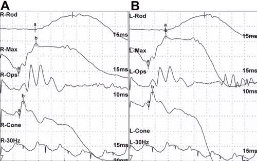
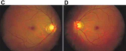
Figure 1. ERG recording and fundus photographs of the proband
A,B: ERG recorded under scotopic and photopic conditions according to ISCEV standards of individual 12 in Figure 2. Panel A shows the right eye and panel B shows the left eye. C,D: Photographs showing typical changes of high myopia, including optic nerve head crescents and "tigroid" appearance of the posterior retina. Panel C shows the right fundus and panel D shows the left fundus of individual 12.