![]() Figure 1 of
Zhou, Mol Vis 2005;
11:414-424.
Figure 1 of
Zhou, Mol Vis 2005;
11:414-424.
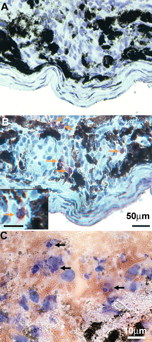
Figure 1. Early neutrophil response during laser induced CNV
A: A laser lesion was stained by a rat IgG2b mAb, an isotype control for anti-Ly-6G. B: Neutrophils, identified by arrows, were detected with anti-Ly-6G mAb in the site of laser injury. Bar in inset represents 20 μm. C: H & E staining of the laser lesion demonstrated the presence of neutrophils (indicated by arrows) identified by their typical morphology. The laser lesions of A through C were at day 3 after laser photocoagulation. D: Kinetics of neutrophil infiltration into the subretinal region following laser injury. The relative quantity of infiltrating neutrophils was calculated based on the anti-Ly-6G fluorescence volume. The bars represent the mean for 10 eyes at each time; the error bars represent the standard deviation.