![]() Figure 4 of
Montiani-Ferreira, Mol Vis 2005;
11:11-27.
Figure 4 of
Montiani-Ferreira, Mol Vis 2005;
11:11-27.
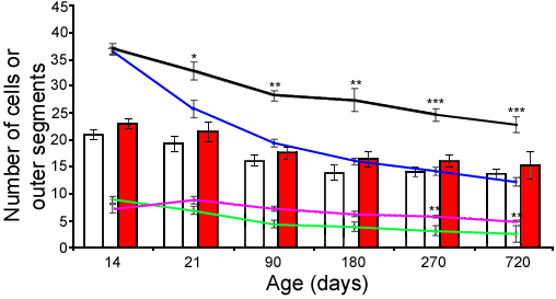
Figure 4. Retinal cell counting
Number of rod OSs and ganglion cells per unit length and number of rows in the INL (average of the six retinal regions demonstrated in Figure 1) in cross sections at 14, 21, 90, 180, 270, and 720 days of age. Note the progressive and significant (p<0.01) overall decrease in number of rod OSs per 200 μm of retinal length in the rge/rge retinas (compare black control line with blue line). At 21 days of age the difference was significant. Additionally, the mean number of rows of nuclei in the INL became significantly smaller in the rge/rge group (compare pink control line to green rge/rge line) at 270 days of age (p<0.0001; 4 rge/rge, 3 controls). The mean number of ganglion cells per 200 μm of retinal length was not statistically significant between the two groups (compare red control columns to white rge/rge columns). A mean number of 4 retinal samples from rge/rge and control birds were analyzed from each age group. Asterisks indicate significance. The single asterisk indicates p<0.01, the double asterisks indicates p<0.001, and the triple asterisks indicates p<0.0001.