![]() Figure 1 of
Montiani-Ferreira, Mol Vis 2005;
11:11-27.
Figure 1 of
Montiani-Ferreira, Mol Vis 2005;
11:11-27.
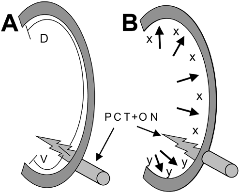
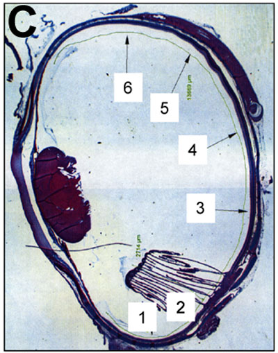
Figure 1. Selection of regions for retinal thickness measurement
Graphical representation of the method used for determining the retinal regions used for retinal thickness measurement. A: Measurements were made of retinal length from the ora serrata; dorsal (D) and ventral (V) to the pecten/optic nerve (PCT+ON). B: The value D was divided by 5, resulting in x and the value V was divided by 3, resulting in y. Starting at the center of PCT+ON, x and y values (dorsally and ventrally, respectively) were consecutively applied to determine the regions to be evaluated for retinal thickness. C: Cross section of a chick eye globe showing the 6 positions at which retinal thickness measurements were made.

