![]() Figure 5 of
Hemati, Mol Vis 2005;
11:1151-1165.
Figure 5 of
Hemati, Mol Vis 2005;
11:1151-1165.
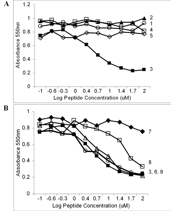
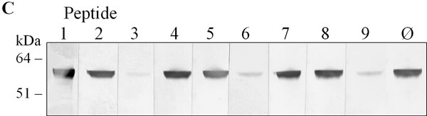
Figure 5. Peptide competition of mAb 8B11 binding
A,B: Competition ELISA of mAb 8B11 binding with synthetic peptides corresponding to sequences in RPE65 Region 2. RPE membranes in microtitre plates were incubated with mAb 8B11 plus peptides at concentration range of 0.1 to 100 μM. A: Competition with peptide 1 are circles, 2 are triangles, 3 are black rectangles, 4 are white rectangles, 5 are rhomboids (sequences shown in Figure 3). B: Competition with derivatives of peptide 3 (black rectangles) made by deleting residues from the amino (peptide 7, rhomboids) or carboxyl (peptide 6, triangles; peptide 8, white rectangles) ends, or both (peptide 9, white rectangles). Data are representative of three independent experiments. C: Western analysis of peptide competition of mAb 8B11 binding. Bovine RPE membrane proteins separated by SDS-PAGE and blotted on nitrocellulose were incubated with mAb 8B11 plus various peptides (100 μM) as shown, and reactivity visualized using alkaline phosphatase coupled anti-mouse IgG.