![]() Figure 4 of
Hemati, Mol Vis 2005;
11:1151-1165.
Figure 4 of
Hemati, Mol Vis 2005;
11:1151-1165.
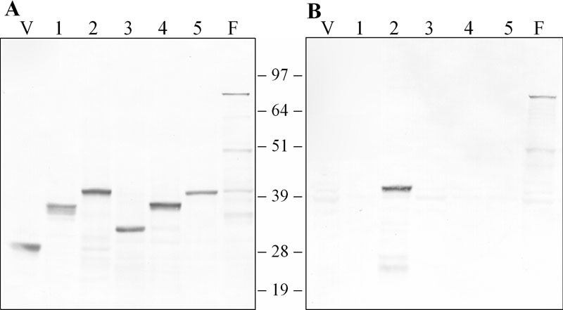
Figure 4. Immunoreactivity of RPE65-fusion proteins
Western analysis of yeast strains transfected with RPE65-pHybLex/Zeo expression constructs encoding RPE65 Regions 1-5 shown in Figure 3. Soluble proteins from cells lysed in 8 M urea and 5% SDS were separated by SDS-PAGE and transferred to nitrocellulose. Blots were probed with anti-LexA antibody (A) or mAb 8B11 (B), and reactivity visualized using alkaline phosphatase coupled anti-mouse IgG. The vector is in the lane labeled "V", the full length RPE65 cDNA is in the lane labeled "F", and regions 1-5 are in the numbered lanes.