![]() Figure 5 of
Mansfield, Mol Vis 2004;
10:728-737.
Figure 5 of
Mansfield, Mol Vis 2004;
10:728-737.
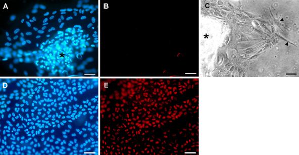
Figure 5. Lack of Pax-6 expression in explants cultured with TGFβ, FGF, and DEX
Explants were cultured with TGFβ then FGF with inclusion of DEX throughout the entire culture period (A,B,C), as described in the legend to Figure 2. They were then photographed by phase contrast microscopy (C) or fixed as whole mounts and processed for immunolocalization of Pax-6 (B) with Hoechst counterstaining of nuclei (A). An explant cultured without addition of growth factors for 7 days and processed in parallel provided a positive control for Pax-6 expression (D, Hoechst dye; E, Pax-6). In TGFβ/FGF/DEX treated explants multilayered plaques formed (A, asterisk indicates plaque); these were associated with a monolayer of cells radiating out from the base of the plaque onto the lens capsule. Pax-6 reactivity was not detectable in either the plaque or the surrounding monolayer of cells (B), whereas cells in the positive control exhibited strong nuclear reactivity for Pax-6 under these localization conditions (D,E). A phase contrast micrograph (C) of an explant analogous to that depicted in A and B shows a monolayer of cells spreading out from the base of a plaque (asterisk); cells at the leading edge have developed lamellae (arrowheads), a characteristic of actively migrating cells. The bar represents 50 μm in A, B, D, and E and 40 μm in C.