![]() Figure 2 of
Mansfield, Mol Vis 2004;
10:728-737.
Figure 2 of
Mansfield, Mol Vis 2004;
10:728-737.
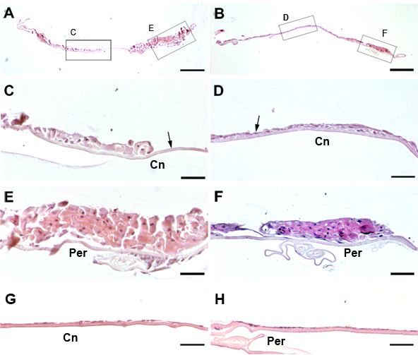
Figure 2. Effect of dexamethasone on cell morphology: hematoxylin and eosin stained sections
Lens epithelial explants were treated with TGFβ then FGF without addition of DEX (A,C,E) or with addition of DEX (B,D,F), as described in the legend to Figure 1, or treated in parallel with DEX alone (G,H). Medium was replaced 5 days after addition of FGF and every 4-6 days thereafter, with re-addition of FGF (A-F) and/or re-addition of DEX (B,D,F-H). At the end of the culture period explants were fixed, embedded and sectioned transversely. Low power images of sections are shown in A and B, with labeled boxes to indicate the regions shown at higher magnification in C,D,E, and F. In TGFβ/FGF treated explants in the absence of DEX, explants exhibited multilayering and extensive enlargement of cells (C,E) and, between cellular aggregates, regions of denuded lens capsule were visible (C, arrow). When DEX was included, multilayering also occurred but there appeared to be less cell enlargement and most of the lens capsule remained covered with cells (B,D,F), although in some regions only a thin monolayer of cells was present (D, arrow). With or without DEX, the explants were thicker in the peripheral region (E,F) than in the central region (C,D). In contrast, explants cultured in parallel with DEX alone generally remained covered with a monolayer of cuboidal cells in both the central (G) and peripheral (H) region, as in the normal lens epithelium. The bar represents 250 μm in A and B and 50 μm in C-H. The peripheral region (Per) and the central region (Cn) of the explants are labeled.