![]() Figure 1 of
Mansfield, Mol Vis 2004;
10:728-737.
Figure 1 of
Mansfield, Mol Vis 2004;
10:728-737.
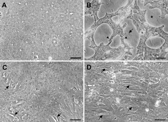
Figure 1. Effect of dexamethasone on cell morphology: phase contrast microscopy
Explants were cultured in control medium (A) or with 50 pg/ml TGFβ2 for 1 day (B-D), then medium was replaced and 20 ng/ml FGF-2 was added to some cultures (C,D). In D only, 100 nM DEX was included on day 0 and day 1. Explants were cultured for a total of 5 (A,B) or 6 (C,D) days, then photographed. Cells in control explants, cultured without growth factors or DEX, tended to remain in cobblestone arrays typical of the normal lens epithelium, as shown in A. Treatment with TGFβ only (B) resulted in extensive cell surface blebbing (arrowheads) associated with loss of cells from the lens capsule (asterisk) and wrinkling of the lens capsule (arrow). Adding FGF on day 1 largely prevented these changes (C,D). Under the latter conditions, in the absence of DEX (C), explants remained well covered with cells, which included occasional spindle-like cells (arrows). However, inclusion of DEX (D) resulted in the formation of numerous parallel arrays of spindle-like cells (arrows) throughout the explant. The bar represents 30 μm in A and B and 75 μm in C and D.