![]() Figure 4 of
Mansfield, Mol Vis 2004;
10:728-737.
Figure 4 of
Mansfield, Mol Vis 2004;
10:728-737.
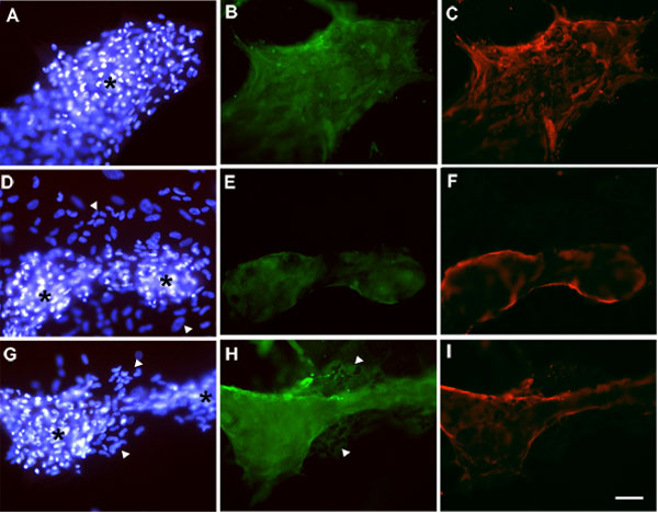
Figure 4. Immunolocalization of α-smooth muscle actin and fibronectin in cell cultures
Lens epithelial explants were cultured with TGFβ then FGF, without DEX (A-C) or with addition of DEX on day 0 and at each change of medium throughout the entire culture period (D-F), as described in the legend to Figure 2. Other explants (G-I) were treated with TGFβ/FGF/DEX as for D-F but DEX treatment was discontinued from day 10 of culture onwards. Explants were fixed as whole mounts and processed for immunolocalization of α-smooth muscle actin (B,E,H; green) and fibronectin (C,F,I; red) by a double labeling technique, with Hoechst counterstaining of nuclei (A,D,G). In all treatment groups, reactivity for both α-smooth muscle actin (B,E,H) and fibronectin (C,F,I) was detected wherever cells had become multilayered and formed into plaques (asterisks in A,D,G). Reactivity for α-smooth muscle actin was relatively weak in plaques in explants treated continuously with DEX (E) but particularly strong in explants in which DEX treatment was discontinued at 10 days (H). Monolayered regions of cells adjacent to plaques were present only in explants that received DEX, whether DEX was present continuously (D, arrowheads) or discontinued at 10 days (G, arrowheads). In the former treatment group, no reactivity for the PCO markers was present in the monolayered cells (E,F), while in the latter group, reactivity for α-smooth muscle actin was readily detectable in these cells (H, arrowheads) but reactivity for fibronectin was not (I). The bar represents 80 μm.