![]() Figure 3 of
Mansfield, Mol Vis 2004;
10:728-737.
Figure 3 of
Mansfield, Mol Vis 2004;
10:728-737.
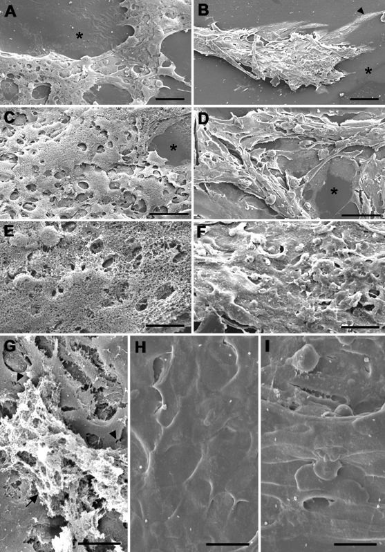
Figure 3. Effect of dexamethasone on cell morphology: SEM
Lens epithelial explants were treated with TGFβ then FGF without addition of DEX (A,C,E,G) or with addition of DEX (B,D,F,H,I), as described in the legend to Figure 2, then processed for SEM. Asterisks indicate regions of denuded lens capsule. In these TGFβ/FGF treated explants, both in the absence of DEX (A) and the presence of DEX (B), thick plaques of cells formed during culture. In the presence of DEX, however, a monolayer of attenuated cells formed, extending from the base of the plaque across the lens capsule (B, arrowhead). The general topography of these explants was also markedly different (C,D). Examination at higher magnification revealed that the explant cultured without DEX was covered with a thick web of ECM-like material, which completely obscured the cellular surface (E). In another TGFβ/FGF treated explant cultured without DEX (G), ECM-like material was present in scattered patches (arrow), and flattened, elongated cells were visible, which exhibited occasional tongue and flap-like, interdigitating processes at their margins (G, arrowheads). In contrast, the DEX treated explant shown in B and D exhibited only very sparse deposition of ECM-like material (F), wh ile extensive regions of another explant in this treatment group appeared completely devoid of surface ECM (H,I). The cells in the DEX treated explants exhibited diverse and unusual morphologies (F,G,H). The bars represent 50 μm in A-D and 20 μm in E-I.