![]() Figure 6 of
Stoilov, Mol Vis 2004;
10:629-636.
Figure 6 of
Stoilov, Mol Vis 2004;
10:629-636.
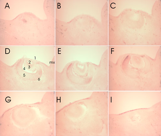
Figure 6. Serial transverse sections through the developing eye of 11.5 dpc embryo
A series of ten 50 μm sections were taken in a nasal to temporal direction through the developing eye of 11.5 dpc embryo. One damaged section located between F and G was not included. Based on the number of sections the diameter of the developing eye at this stage is estimated to be approximately 500 μm. The plane of the sections was selected to slice through both ocular expression domains and the maxillary component of the first branchial arch (mx) as seen in Figure 5B. Corneal ectoderm (epithelium) (1), cavity of the lens vesicle (2), lens epithelium (3), future hyaloid cavity (4), inner (neural) layer of the optic cup representing the future nervous layer of the retina (5), and the outer layer of the optic cup representing the future pigmented layer of the retina (6) are numbered. The maxillary component of the first branchial arch (mx) is labeled.