![]() Figure 5 of
Mansfield, Mol Vis 2004;
10:521-532.
Figure 5 of
Mansfield, Mol Vis 2004;
10:521-532.
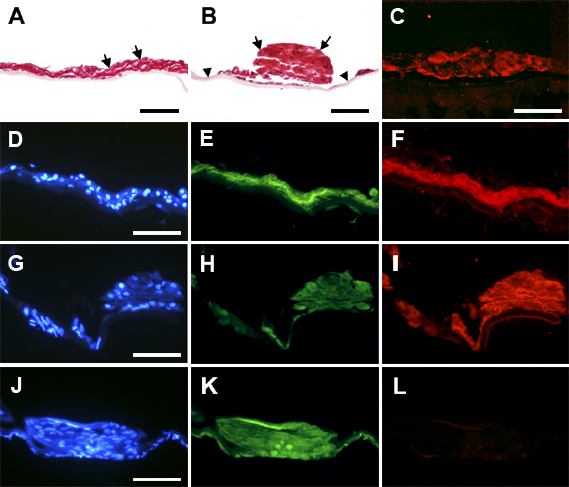
Figure 5. Immunolocalization of various markers in sections of lens epithelial explants cultured with TGFβ then FGF
Lens epithelial explants were cultured with TGFβ2 at a concentration of 50 pg/ml (A,C,D-F) or 100 pg/ml (B,G-L) for one day, then with 100 ng/ml FGF-2 for 21 days, as described in the Legend of Figure 4. At the end of the culture period, explants were fixed, embedded in paraffin and sectioned. Sections were stained with hematoxylin-eosin (A,B) or processed for immunolocalization of collagen type I (C), or αSMA (E,H,K) and β-crystallin (F,I,L), with Hoechst counterstaining of nuclei (D,G,J). In D-L, each row presents images of the same region. G-I and J-L are from different regions of the same plaque. All images are arranged capsule surface downwards. In the explant shown in A, a thick multilayer of cells covered the entire lens capsule, instead of the monolayer present in control explants and the normal lens. In the explant shown in B, the cells mounded up into a plaque leaving regions of denuded capsule (arrowheads). In both cases, nuclei were sparse in some regions (A, B, arrows), indicating cell enlargement, and virtually all cells showed reactivity for type I collagen (as seen in C) and αSMA (E,H,K). In the explant that remained covered with multilayered cells, strong reactivity for β-crystallin was detected throughout the entire explant (F). In the explant that exhibited plaque formation, reactivity for β-crystallin was particularly strong in one region of a plaque (I) but virtually absent from another (L). The bar represents 85 μm.