![]() Figure 4 of
Mansfield, Mol Vis 2004;
10:521-532.
Figure 4 of
Mansfield, Mol Vis 2004;
10:521-532.
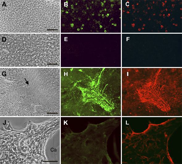
Figure 4. Immunolocalization of αSMA and fibronectin in explants cultured with TGFβ then FGF
Lens epithelial explants were cultured for one day without TGFβ2 (A-F) or with 50 pg/ml TGFβ (G-L) then medium was replaced and FGF-2 was added to D-L at a final concentration of 20 ng/ml. Explants were cultured for a total of 11 days (A-I) or 31 days (J-L) with replacement of medium every fifth day, and re-addition of FGF (D-L only). They were then fixed and processed as whole mounts for immunolocalization of αSMA (B,E,H,K) or fibronectin (C,F,I,L) using a double labeling technique. Phase contrast images of corresponding regions are also included in each row (A,D,G,J). Explants cultured for 11 days without addition of growth factors retained typical epithelial morphology (A) but scattered cells showed reactivity for αSMA (B) and fibronectin (C). Cells in corresponding explants cultured with FGF alone were arrayed more irregularly (D) and no specific reactivity for αSMA (E) and fibronectin (F) was detectable. TGFβ/FGF treated explants cultured in parallel contained abundant tightly packed cells with occasional distinctive conglomerates of cells (G, arrow). The latter exhibited strong reactivity for both αSMA (H) and fibronectin (I) and many cells in this region were spindle shaped (G-I). By 31 days of culture (J-L), cells in the TGFβ/FGF treated explant depicted gathered up into a thick mound, leaving the surrounding lens capsule (Ca) denuded. Reactivity for αSMA (K) and fibronectin (L) was detectable. The bar represents 75 μm in A-F and J-L and 150 μm in G-I.