![]() Figure 2 of
Harrington, Mol Vis 2004;
10:476-489.
Figure 2 of
Harrington, Mol Vis 2004;
10:476-489.
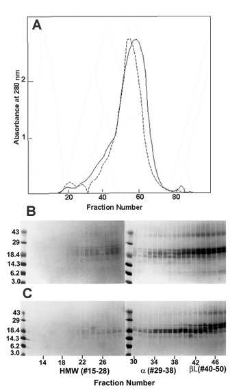
Figure 2. Isolation and identification of WS-HMW protein fractions from normal and cataractous human lenses
This figure shows separation of the HMW-protein and α-crystallin fractions from the WS proteins of human lenses following size-exclusion chromatography. Isolation of WS-HMW protein fraction following size exclusion Agarose A 5 m chromatography from WS protein fraction of normal and cataractous lenses (both from 50-60 year old donors). The HMW protein eluted as a void volume peak (fractions 15-28). A: Protein elution profile at 280 nm of WS-HMW proteins from 50-60 year old normal (solid line) and cataractous (dashed line) lenses during size exclusion Agarose A 5m chromatography. B: Identification of WS-HMW protein fractions (fractions 15-28) and WS-α-crystallin protein fractions (fractions 29-38) by SDS-PAGE using a 15% polyacrylamide gel.