![]() Figure 5 of
Fautsch, Mol Vis 2004;
10:417-425.
Figure 5 of
Fautsch, Mol Vis 2004;
10:417-425.
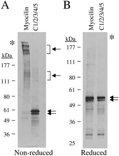
Figure 5. Myocilin forms disulfide bonds in vitro
DNA templates containing open reading frames of myocilin or myocilin containing substitutions of alanine for all five cysteines (C1/2/3/4/5) were made (see Figure 3 for schematic diagram of these constructs). A: 35S-labeled proteins obtained from in vitro transcription/translation reactions (40 μl) from each construct were immunoprecipitated with hemagglutinin polyclonal antibody. Immunoprecipitated products were bound to protein A-sepharose, washed in RIPA buffer, and separated on 7% denatured, non-reduced SDS-PAGE gels. Gels were transferred to PVDF membrane followed by autoradiography. B: 35S-labeled proteins obtained from in vitro transcription/translation reactions (10 μl, not immunoprecipitated) from each construct were separated on 11% denatured, reduced SDS-PAGE gels. Gels were transferred to PVDF membrane followed by autoradiography. Arrows indicate prominent myocilin complexes. Asterisks indicate border between stacking and resolving gel.