![]() Figure 3 of
Fautsch, Mol Vis 2004;
10:417-425.
Figure 3 of
Fautsch, Mol Vis 2004;
10:417-425.
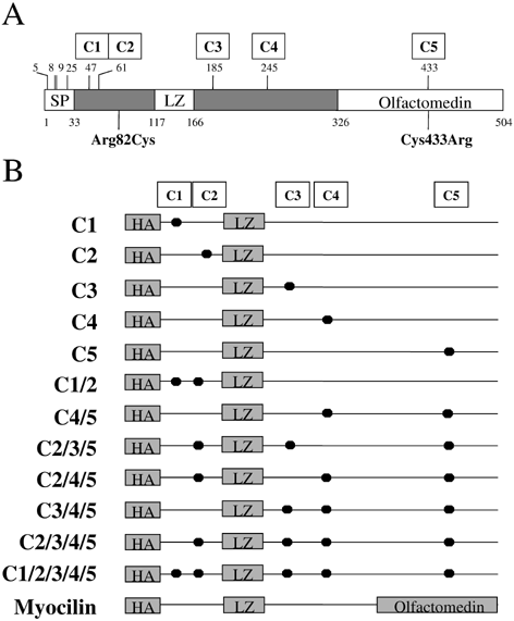
Figure 3. Schematic diagram of myocilin
A: Top numbers represent location of cysteine amino acids found in human myocilin. The clinical mutation Arg82Cys represents a cysteine substitution at amino acid 82 for arginine. The clinical mutation Cys433Arg represents a arginine substitution at amino acid 433 for cysteine. B: Schematic diagram of fusion proteins used for in vitro disulfide bond studies. Dots indicate where a cysteine amino acid was changed to alanine. C1 through C5 represent amino acids 47, 61, 185, 245, and 433, respectively. Abbreviations are denoted as follows: HA, N-terminal tag containing an epitope for a hemagglutinin polyclonal antibody; SP, signal peptide sequence; LZ, leucine zipper motif.
