![]() Figure 6 of
Liou, Mol Vis 2004;
10:383-391.
Figure 6 of
Liou, Mol Vis 2004;
10:383-391.
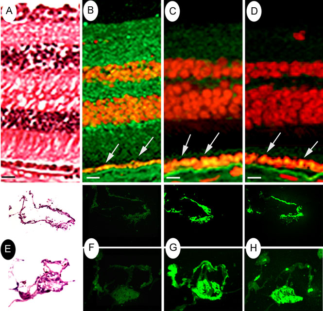
Figure 6. Immunolocalization of conventional APC and BS-APC in the epiretinal membrane
Laser-scanning confocal microscopic immunolocalization of conventional APC, BS-APC, and pan-cytokeratin is shown in human retina (A-D) and two different regions of an ERM from one patient (P5). E-H: Serial vertical sections (x and z) of formaldehyde fixed retina sections or cryosectioned ERM incubated with different antibodies and with Oregon Green-conjugated secondary antibodies. A,E: H & E stained. B,F: anti-BS-APC. C,G: anti-exon 1 (Ab-1). D,H: anti-pan-cytokeratin. Cell nuclei were stained with propidium iodide. Arrows indicate RPE monolayer, which is seen as an orange hue. Bar represents 10 μm.