![]() Figure 4 of
Liou, Mol Vis 2004;
10:383-391.
Figure 4 of
Liou, Mol Vis 2004;
10:383-391.
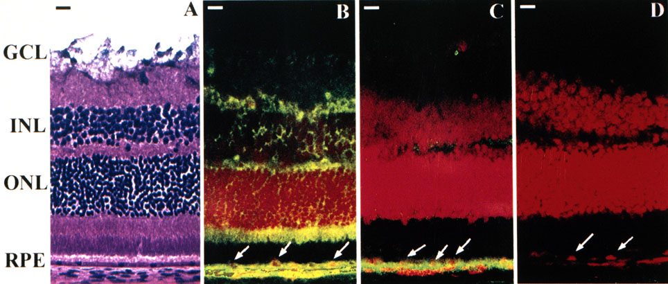
Figure 4. Retinal immunolocalization of conventional APC and BS-APC
Images are of laser scanning confocal microscopic immunolocalization of conventional APC and BS-APC in mouse retina. A: H & E stained cryosection depicting ganglion (GCL), inner and outer nuclear layer (INL and ONL), and RPE cells. B-D: Vertical sections (x and z) taken of cryosection of mouse eye incubated with different antibodies against APC and Oregon Green-conjugated secondary antibodies. B: Anti-BS. C: Anti-exon 1 (Ab-1). D: Normal serum. Cell nuclei were stained with propidium iodide. Arrows indicate RPE cell nuclei. Bar represents 10 μm.