![]() Figure 3 of
Ruiz-Ederra, Mol Vis 2004;
10:83-92.
Figure 3 of
Ruiz-Ederra, Mol Vis 2004;
10:83-92.
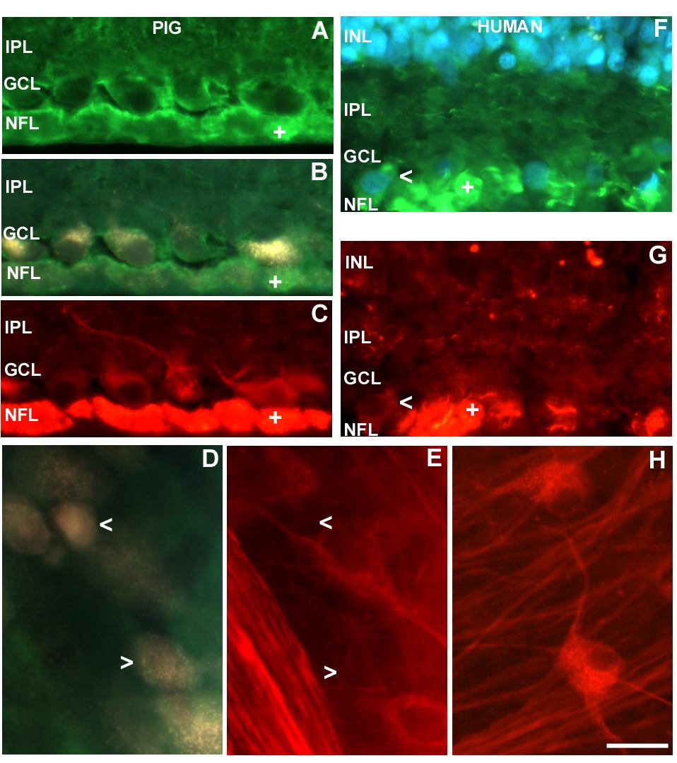
Figure 3. Distribution of NF-Hp and NF-M
Distribution of NF-Hp (green) and NF-M (red) in double labeled pig and human retinal sections and flatmounts (A, B, and C). Sections from pig retina immunostained with NF-H (A), back filled with fluorogold (B), and NF-M (C). A: As shown in Figure 2A, NF-Hp is located in axon bundles (+) and dendrites of retinal ganglion cells (RGCs). C: The same RGCs possess NF-M distributed within the cytoplasm and dendrites and in axon bundles (+). D and E: Flat mounts of pig peripheral retina back filled with fluorogold (D) and then immunostained with NF-M (E), showing immunolabeling in RGC processes and somas. Open arrowheads point to RGCs that are not immunolabeled for NF-M. F and G: Section from human peripheral retina immunostained with NF-Hp (nuclei labeled with DAPI; F), and NF-M (G). F: As shown in Figure 2E this antibody binds axon bundles in the nerve fiber layer (NFL; +), the soma of some RGCs (open arrowhead) and many processes in the inner plexiform layer (IPL). G: NF-M is present in somas (open arrowhead), axons and processes of RGCs. H: Flatmount from human retina immunostained with NF-M. Processes and somas of RGCs can be seen immunolabeled with this antibody. The scale bar for A, B, C, and F represents 20 μm. The scale bar for D, E, and G represents 40 μm. The nerve fiber layer (NFL), ganglion cell layer (GCL), inner plexiform layer (IPL), and inner nuclear layer (INL) are labeled in the images.