![]() Figure 2 of
Ruiz-Ederra, Mol Vis 2004;
10:83-92.
Figure 2 of
Ruiz-Ederra, Mol Vis 2004;
10:83-92.
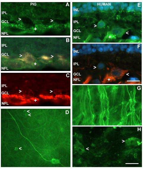
Figure 2. Distribution of NF-Hp and NF-H
Distribution of NF-Hp (green) and NF-H (red), in double labeled pig and human retinal sections and flatmounts. A, B, and C: Section showing the inner layers of the pig retina double-immunolabeled with NF-Hp (A), NF-H (C), and backfilled with fluorogold (B). The open arrowheads point to RGCs that present staining mainly in axons and dendrites with anti-NF-Hp (A) and in axon, dendrites and cytoplasm with anti-NF-H (C). D: Flat mount from peripheral retina of pig immunostained with NF-Hp. Together with the axons, a regular mosaic of small RGCs (open arrowheads) surrounding a large RGC is visible. E and F: Sections from human peripheral retina double-immunolabeled with NF-Hp (E) and NF-H (F). Nuclei were labeled with DAPI. Both NF antibodies bind axon bundles in the NFL (+), RGC somata and dendrites in the inner plexiform layer (IPL). A RGC immunopositive for NF-Hp but unlabeled for NF-H can be seen (left open arrowhead). G: Flatmount from human central retina immunostained with NF-Hp. Only robustly stained axons can be seen. H: Flatmount from human peripheral retina immunostained with NF-H, label visible mainly in the cytoplasm of RGCs (open arrowheads). The scale bar represents 20 μm for all pictures except in D, where it represents 40 μm. The nerve fiber layer (NFL), ganglion cell layer (GCL), inner plexiform layer (IPL), and inner nuclear layer (INL) are labeled in the images.