Figure 6. Antioxidant treatment suppresses NaIO3-induced cytotoxicity and restore gene expression toward baseline in induced pluripotent stem cell-retinal pigment epithelium
cells. Effect of NaIO3 on hiPSC-RPE cell viability in the presence or absence of a broad redox-active antioxidant. Cells were pre-treated with Mn-TM-2-PyP
(0.005%) for 24 h and during NaIO3 exposure. A: Cell viability measured by intracellular resazurin reduction assay, expressed as a percentage relative to untreated control
cells. The half-maximal inhibitory concentration (IC50) indicates the NaIO3 concentration causing 50% cell death. B: Cell viability assessed by lactate dehydrogenase (LDH) release. The half-maximal effective concentration (EC50) represents the NaIO3 concentration at which 50% of maximal LDH release was observed. Gene expression changes in the presence or absence of antioxidant
at NaIO3 concentrations of 4–6 mM. Expression of C: Vascular endothelial growth factor A (VEGFA), D: Hypoxia-inducible factor 1-alpha (HIF1A), E: Thioredoxin (TXN), F: Thioredoxin interacting protein (TXNIP). G: Gamma-glutamylcyclotransferase 1 (CHAC1) and H: Cystine/glutamate antiporter (SLC7A11) were normalized to control condition without antioxidant. Statistical analysis was performed using two-way ANOVA with Šidák’s
multiple comparison test. *p < 0.05, **p < 0.01, ***p < 0.001, ****p < 0.0001. Each technical replicate is shown as a single
data point with mean ± SD (n = 2).
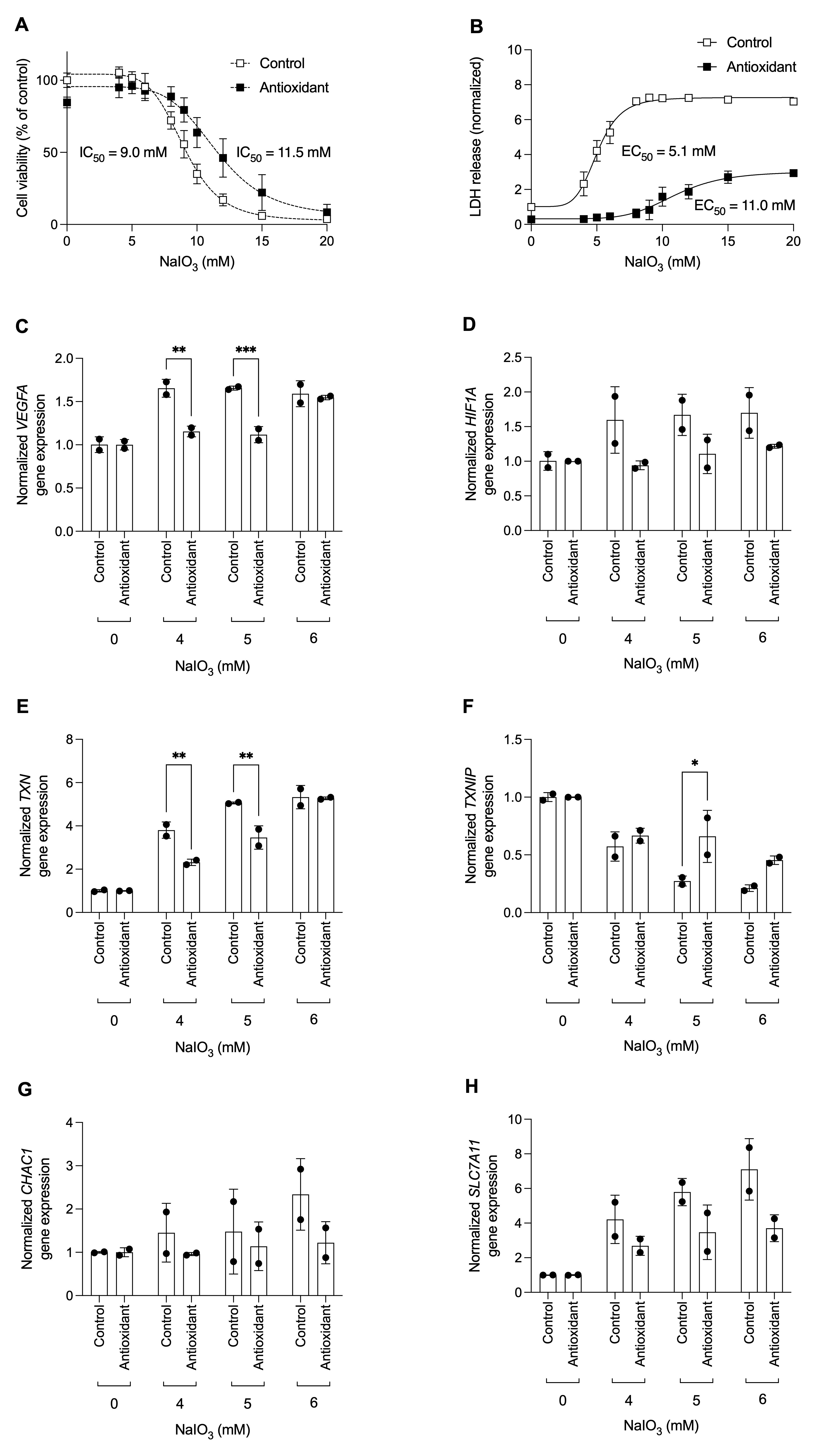
 Figure 6 of
Kolehmainen, Mol Vis 2026; 32:421-433.