Figure 4. Cellular responses of human induced pluripotent stem cell-derived retinal pigment epithelium cells to 24 h NaIO3- and tBHP-induced oxidative stress. A: Cell viability measured as intracellular reduction of resazurin and reactive oxygen species (ROS) formation after NaIO3-induction. Cell viability is shown as a percentage of live cells compared to control cells. The half-maximal inhibitory concentration
(IC50) represents the concentration with which 50% of the cells died. B: Lactate dehydrogenase (LDH) and vascular endothelial growth factor A (VEGFA) release after NaIO3-induction. The half-maximal effective concentration (EC50) represents the concentration of NaIO3 with which half of LDH maximum release was reached. C: Cell viability and ROS formation after tBHP-induction. D: LDH and VEGFA release after tBHP-induction. ROS and LDH levels were normalized control condition (0 mM). VEGFA release is
shown as ng/ml. All data are shown as mean ± SD (n = 3).
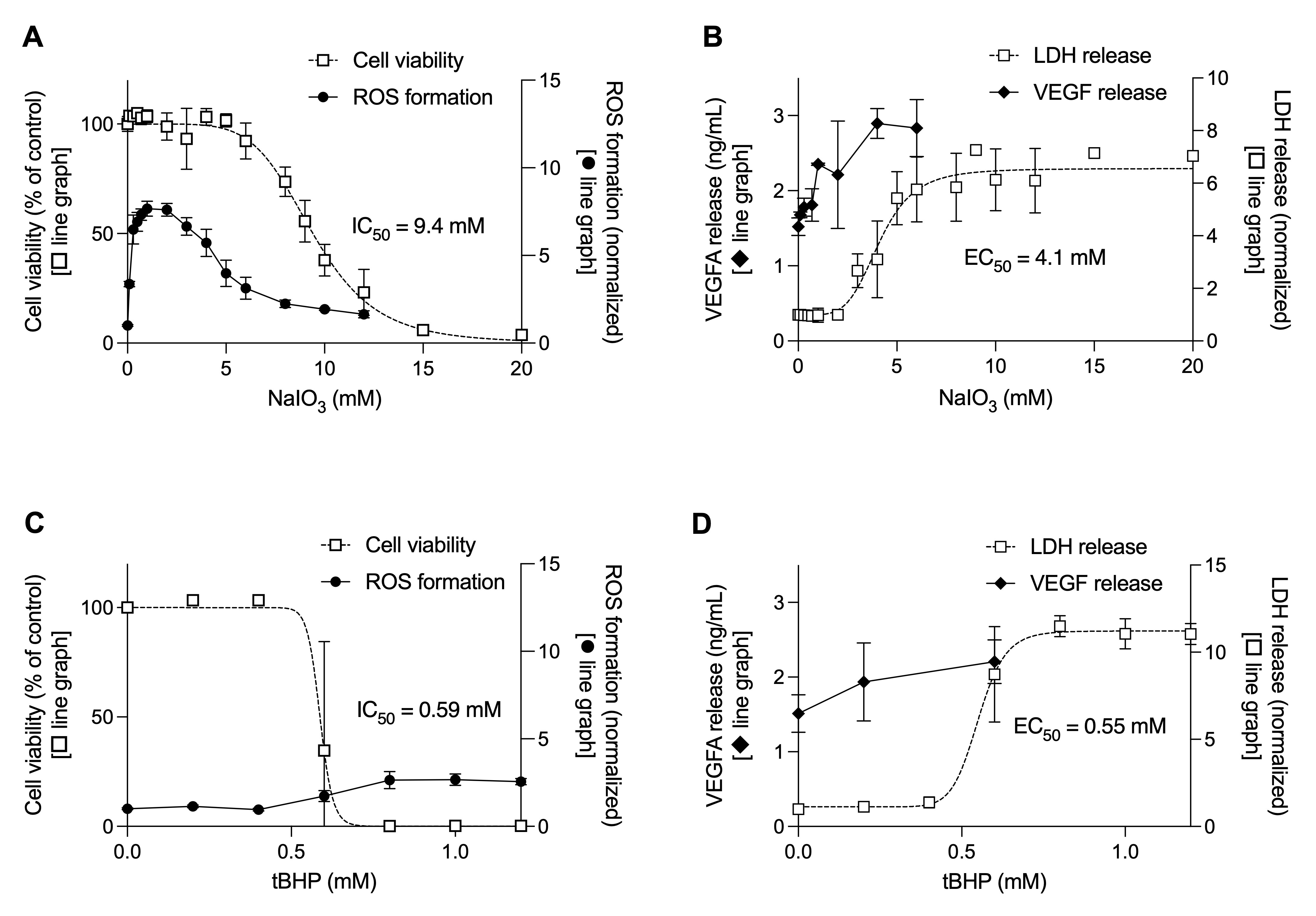
 Figure 4 of
Kolehmainen, Mol Vis 2026; 32:421-433.