Figure 3. Melanosome density and maturation in human induced pluripotent stem cell-derived retinal pigment epithelium cells. A: Representative transmission electron microscopy image of human induced pluripotent stem cell-derived retinal pigment epithelium
(hiPSC-RPE) cells containing immature melanosomes at stages I (yellow arrow) and II (blue arrow), and mature melanosomes at
stages III (orange arrow) and IV (black arrow). Scale bar 500 nm. B: Number of melanosomes per hiPSC-RPE cell according to maturation stage. Data are presented as mean ± SD (n =24).
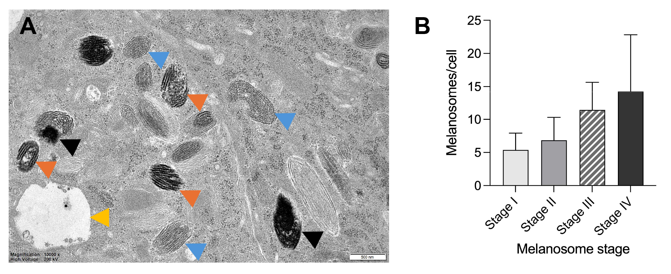
 Figure 3 of
Kolehmainen, Mol Vis 2026; 32:421-433.