Figure 2. Assessment of maturity in human induced pluripotent stem cell-derived retinal pigment epithelium monolayers. Representative
immunofluorescence images of human induced pluripotent stem cell-derived retinal pigment epithelium showing A: occludin, B: primary cilia (ARL13B), C: merged staining of occludin (green), primary cilia (grey), and DAPI (blue), and D: expression of the RPE maturity marker RPE65. Scale bars: 5 µm (A–C) and 100 µm (D).
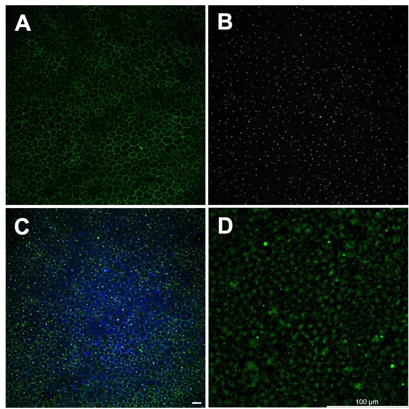
 Figure 2 of
Kolehmainen, Mol Vis 2026; 32:421-433.