Figure 1. Characterization of tight junction protein expression, localization, and morphology in human induced pluripotent stem cell-derived
retinal pigment epithelium. Representative immunofluorescence images of A: zonula occludens-1 (ZO-1) and B: occludin expression in human induced pluripotent stem cell-derived retinal pigment epithelium (hiPSC-RPE) monolayers. Representative
transmission electron microscopy images showing C: vertical and D: horizontal cross-sections of the hiPSC-RPE monolayer. Scale bars: 50 µm (A, B) and 5 µm (C, D).
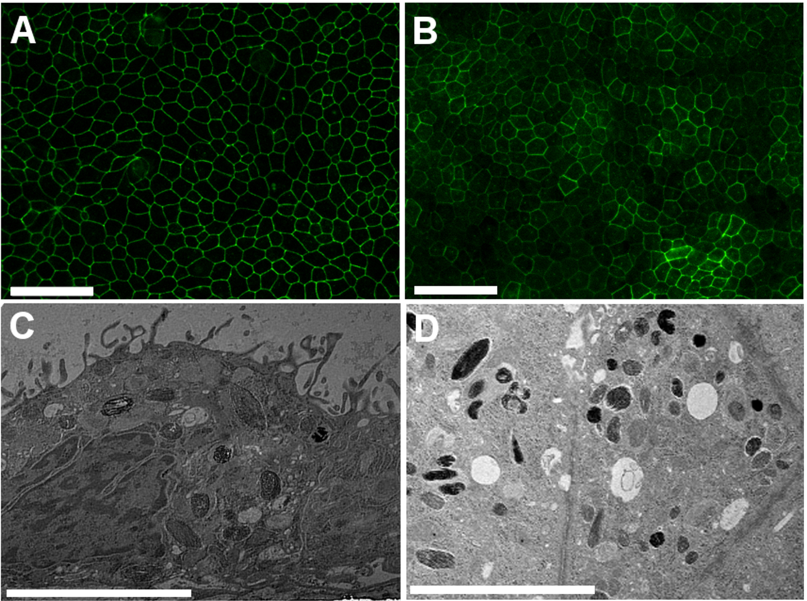
 Figure 1 of
Kolehmainen, Mol Vis 2026; 32:421-433.