![]() Figure 1 of
Haque, Mol Vis 2003;
9:52-59.
Figure 1 of
Haque, Mol Vis 2003;
9:52-59.
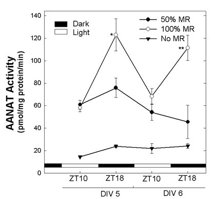
Figure 1. Daily rhythm of AANAT activity in chicken retinal cell cultures
Cultures were prepared from embryonic day 6 retinas. Cells were incubated under a 12 h light:12 h dark (LD) cycle of illumination (light on at ZT 0). After incubation for 24 h at 37 °C on LD, 5μM NBTI was added in the medium and then cells were kept at 40 °C on the same LD cycle for five more days. On day 4 in vitro, one set of the dishes had half of the total medium volume (3 of 6 ml) removed and replaced with 3 ml of medium containing 1% fetal bovine serum and 5 μM NBTI (50% medium replaced [MR]). Another set of dishes had all of the medium replaced with 6 ml of fresh medium containing 1% serum and 5 μM NBTI (100% MR). The third set of dishes was undisturbed and remained in the original medium (No MR). Cells were harvested on days 5 and 6 in vitro (DIV 5, 6) at ZT 10 and ZT 18. The horizontal bars on the bottom of the graph represent the periods of light (open bar) and darkness (filled bar). Data are shown as means with the error bars representing the SEM (N=3-5). AANAT activity increased significantly from ZT 10 to ZT 18 on DIV 5 (p=0.005) and on DIV 6 (p=0.018).
