![]() Figure 6 of
Chen, Mol Vis 2003;
9:735-746.
Figure 6 of
Chen, Mol Vis 2003;
9:735-746.
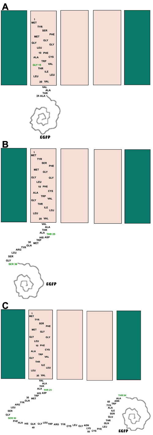
Figure 6. Topology of MP19G truncations in the cell membrane
A: Schematic of a proposed model of MP19-25G integrating into the cell membrane. B: Schematic of a proposed model of MP19-36G integrating into the cell membrane. C: Schematic of a proposed model of MP19-64G integrating into the cell membrane. The globular drawing labeled EGFP represents the EGFP protein fused to the COOH-terminal end of each MP19 truncation.