![]() Figure 4 of
Chen, Mol Vis 2003;
9:735-746.
Figure 4 of
Chen, Mol Vis 2003;
9:735-746.
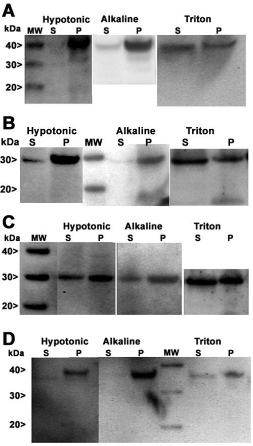
Figure 4. Western analysis of membrane fractions
MP19G- (A), MP19-25G- (B), MP19-36G- (C), and MP19-64G-TRex-293 (D) stable cell lines were grown to confluence in 60 mm dishes and the synthesis of fusion protein induced with tetracycline. After three days of induction, the cells were prepared for analysis of membrane associated protein (Hypotonic lysis), integral membrane protein (Alkaline treated membranes), and lipid raft protein (Triton treated). Samples were subjected to SDS-PAGE under reducing conditions and transferred to nitrocellulose membranes. Membrane were then washed, blocked, incubated with primary antibody (anti-EGFP), washed again, and incubated with secondary antibody conjugated to alkaline phosphatase. The WesternBreeze chemiluminescent immunodetection system (Invitrogen) was used for immunodetection of EGFP fusion protein. MW refers to the molecular weight standards. S refers to the soluble protein fraction and P refers to the pellet (insoluble) protein fraction. MW indicates the molecular weight standard ranging from 20 to 40 kDa.