![]() Figure 4 of
Vinader, Mol Vis 2003;
9:723-729.
Figure 4 of
Vinader, Mol Vis 2003;
9:723-729.
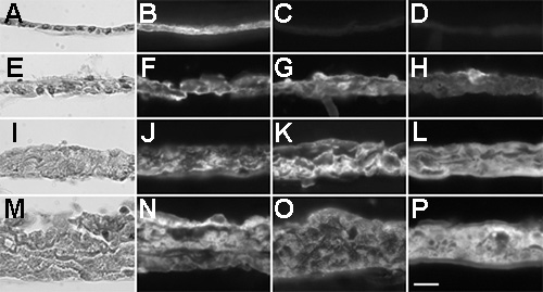
Figure 4. Morphology of and immunofluorescence localization of crystallins in explants cultured for 13 days
Explants were fixed, embedded in paraffin wax, and serial sections cut at 5 μm. One section of every five was mounted and stained with hematoxylin and eosin or used for immunofluorescence staining. Explants were cultured in the absence of growth factors (A-D), with PDGF-AA (E-H), with FGF-2 (I-L), or in the presence of PDGF-AA and FGF-2 together (M-P). Sections from the same explant were stained with hematoxylin-eosin (A,E,I,M) or immunolabeled for αA-crystallin (B,F,J,N), βB2-crystallin (C,G,K,O), or γS-crystallin (D,H,L,P). The scale bar represents 20 μm.