![]() Figure 3 of
Vinader, Mol Vis 2003;
9:723-729.
Figure 3 of
Vinader, Mol Vis 2003;
9:723-729.
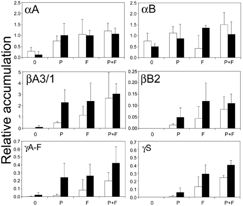
Figure 3. Quantification by dot blot analysis of the accumulation of crystallins in the presence of PDGF-AA with or without FGF-2
Accumulation of αA-, αB-, βA3/1-, βB2-, γC, and γS-crystallin in the absence of growth factors (0), with PDGF-AA (P), with FGF-2 (F), or in the presence of PDGF-AA and FGF-2 together (P+F) during a culture period of 8 (white) or 13 days (black). Samples were spotted for dot blots experiments as described in Methods. The relative intensity of stain of the dots was determined using an imaging densitometer. The data represent the average of three independent experiments and the bar represents the standard deviation. Explant crystallin content was related to a standard range of water-soluble protein extracted from newborn rat lens fiber cells, included in every blot as a crystallin staining control. Crystallin levels are shown relative to the intensity of 100 ng of lens fiber cells loaded in every blot as a staining control.