![]() Figure 2 of
Vinader, Mol Vis 2003;
9:723-729.
Figure 2 of
Vinader, Mol Vis 2003;
9:723-729.
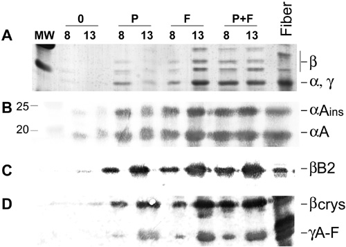
Figure 2. SDS-PAGE and immunoblot analysis of crystallin expression in cultured lens cells in the presence of PDGF-AA and/or FGF-2
Rat lens epithelial explants were cultured in the absence of growth factors (0), with PDGF-AA (P), with FGF-2 (F), or in the presence of PDGF-AA and FGF-2 together (P+F) and harvested after 8 or 13 days of culture. Samples were separated on SDS-PAGE and gels were either silver stained or western blotted as described in Materials and Methods. A: Silver stained gel of explant samples, cultured for the indicated number of days with PDGF-AA and/or FGF-2. MW indicates the lane loaded with molecular weight markers; their molecular weight in kD is indicated next to the band. Fiber marks the lane loaded with a standard amount of water-soluble newborn lens fiber cell protein. An equal fraction of every explant was loaded in each lane. B-D: Western blots of duplicate gels as in A stained with antibodies raised against αA- (the larger splice variant αAins-crystallin found in rodents is also stained, B), βB2- (C) or β-high-crystallin fraction, and γC-crystallin (D). Only the relevant parts of the western blots are shown.