![]() Figure 1 of
Vinader, Mol Vis 2003;
9:723-729.
Figure 1 of
Vinader, Mol Vis 2003;
9:723-729.
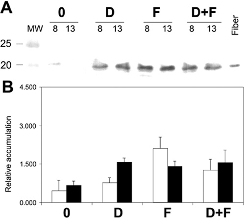
Figure 1. Western blot and quantification of the accumulation of αB-crystallin in rat lens epithelial cells in the presence of dexamethasone and/or FGF-2
Epithelial cells were cultured in the absence of growth factors (0), with dexamethasone (D), with FGF-2 (F), or in the presence of dexamethasone and FGF-2 together (D+F) and harvested after 8 or 13 days of culture. A: Western blot stained for αB-crystallin. B: Quantification of the accumulation of αB-crystallin. Samples were spotted for dot blot experiments as described in Methods. The relative intensity of the stain of the dots was determined using an imaging densitometer. The data represent the average of three independent experiments and the bar represents the standard deviation. Crystallin levels are shown relative to the intensity of 100 ng of lens fiber cells loaded in every blot as a staining control.