![]() Figure 1 of
Rhee, Mol Vis 2003;
9:715-722.
Figure 1 of
Rhee, Mol Vis 2003;
9:715-722.
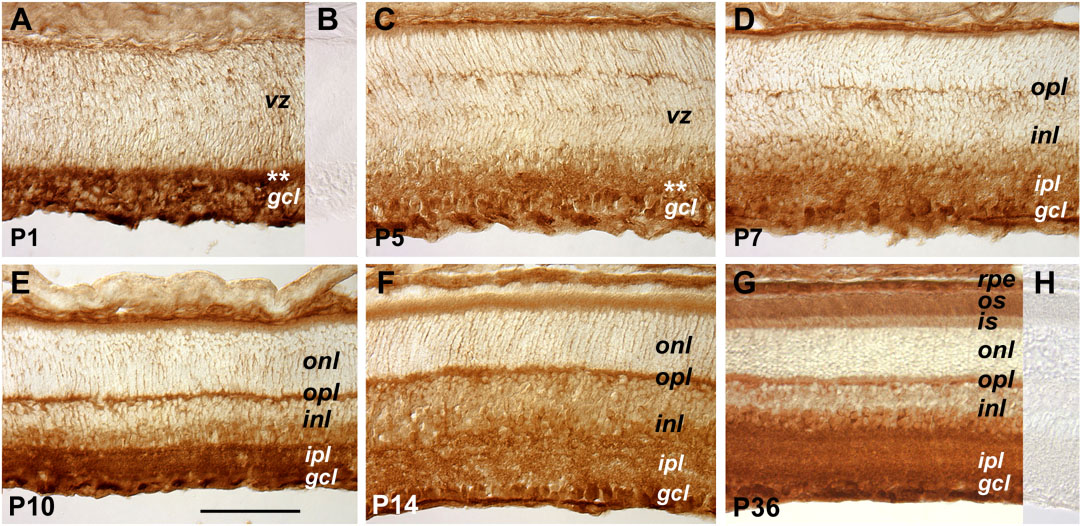
Figure 1. Expression patterns of the gp130 receptor
Cryosections of retinas from different postnatal ages immunohistochemically stained for gp130 are shown. Panels B and H were derived from the same developmental stages as panels A and G, respectively. The primary antibodies used to stain the section shown in panel B were preincubated with the antigen peptide. The staining of panel H was performed without primary antibodies. The asterisks in panels A and C indicate the position of the developing inner plexiform layer at P1 and P5, respectively. The scale bar shown in panel E represents 100 μm for all panels. The meanings of the abbreviations referred to in this figure are: gcl (ganglion cell layer), inl (inner nuclear layer), ipl (inner plexiform layer), is (inner segment), onl (outer nuclear layer), opl (outer plexiform layer), os (outer segment), rpe (retinal pigmented epithelium), vz (ventricular zone).