![]() Figure 4 of
Wang, Mol Vis 2003;
9:701-709.
Figure 4 of
Wang, Mol Vis 2003;
9:701-709.
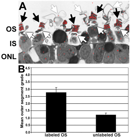
Figure 4. Reversibility of outer segment membrane support
A: Autoradiogram of Xenopus laevis RPE-deprived retina exposed to 5x10⁻⁵ M IPTG and 3H-leucine for two days followed by non-supplemented medium for an additional two days. Red dots indicate the localization of the incorporated 3H-leucine. The outer segment (OS), inner segment (IS), and the outer nuclear layer (ONL) are also labeled in the micrograph. B: Summary graph of outer segment organization during the early and latter portions of the experiment. The black arrows indicate the outer segment membranes containing silver grains that were elaborated during the early part of the culture paradigm, while the white arrows indicate the membranes that do not contain silver grains and were elaborated during the latter part of the experiment.