![]() Figure 2 of
Wang, Mol Vis 2003;
9:701-709.
Figure 2 of
Wang, Mol Vis 2003;
9:701-709.
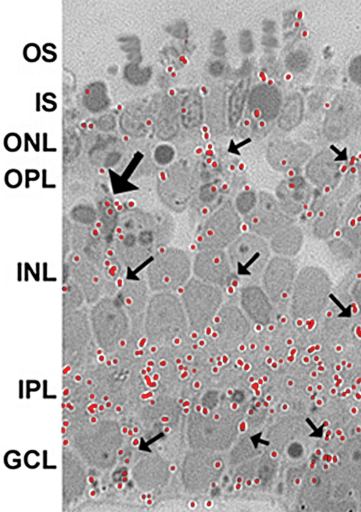
Figure 2. Fate of galactose in the retina
Light microscopic autoradiogram showing incorporation of 3H-galactose in Xenopus laevis retina after three days of culture. The red dots indicate the localization of the incorporated 3H-galactose. The outer segment (OS), inner segment (IS), outer nuclear layer (ONL), outer plexiform layer (OPL), inner nuclear layer (INL), inner plexiform layer (IPL), and the ganglion cell layer (GCL) are also labeled in the micrograph. The small arrows indicate the radial pattern of silver grains throughout the retina while the large arrow indicates a cluster of silver grains over a Müller cell nucleus.