![]() Figure 1 of
Wang, Mol Vis 2003;
9:701-709.
Figure 1 of
Wang, Mol Vis 2003;
9:701-709.
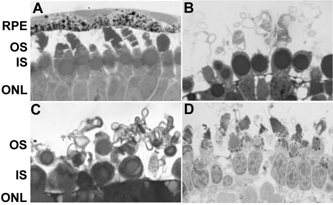
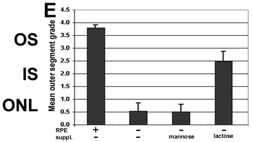
Figure 1. Photoreceptor morphology and outer segment grading
Intact retinas were removed from stage 33/34 Xenopus laevis tadpoles and placed into culture in Niu-Twitty for 3 days. A: RPE-supported retina; B: RPE-deprived retina; C: Mannose-exposed RPE-deprived retina (5x10⁻³ M); D: lactose-exposed RPE-deprived retina (5x10⁻³ M); E: Summary graph of outer segment organization in A-D. The outer segment (OS), inner segment (IS), outer nuclear layer (ONL) are also labeled in the micrograph.