![]() Figure 9 of
Sherry, Mol Vis 2003;
9:673-688.
Figure 9 of
Sherry, Mol Vis 2003;
9:673-688.
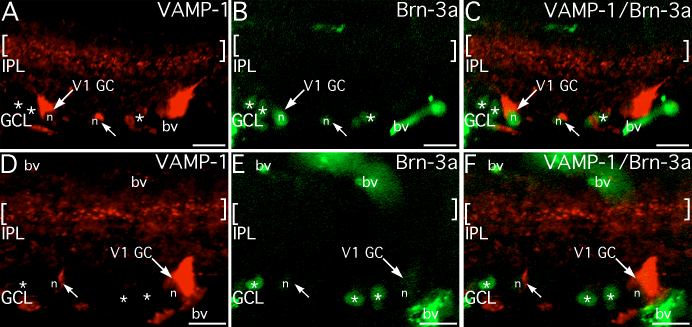
Figure 9. Brn-3a labeling varies among ganglion cells
VAMP-1 and Brn-3a localize to separate cellular compartments: VAMP-1 is restricted to the cytoplasm; Brn-3a is restricted to the nucleus. When co-expressed the two markers are present in the same cell but show non-overlapping distributions within the cell. A-C: A large VAMP-1-immunoreactive ganglion cell (V1 GC) shows particularly strong Brn-3a labeling in its nucleus (n). A small VAMP-1-immunoreactive cell (arrow) that shows only weak Brn-3a labeling in its nucleus (n) is also visible. Several nearby ganglion cells identified by nuclei that are positive for Brn-3a (*) do not show VAMP-1 labeling. A second large VAMP-1-immunoreactive ganglion cell is present in the ganglion cell layer (GCL) at the right, but its nucleus is not visible. D-F: A large ganglion cell at the right (V1 GC) and a small cell at the left (arrow) show labeling for VAMP-1, but neither cell's nucleus (n) shows labeling for Brn-3a. Three nearby ganglion cells do show Brn-3a labeling in their nuclei (*), but do not show labeling for VAMP-1. Brackets denote the VAMP-1-immunoreactive band in the mid-IPL. Blood vessels (bv) show non-specific labeling. Scale bars represent 20 μm.