![]() Figure 8 of
Sherry, Mol Vis 2003;
9:673-688.
Figure 8 of
Sherry, Mol Vis 2003;
9:673-688.
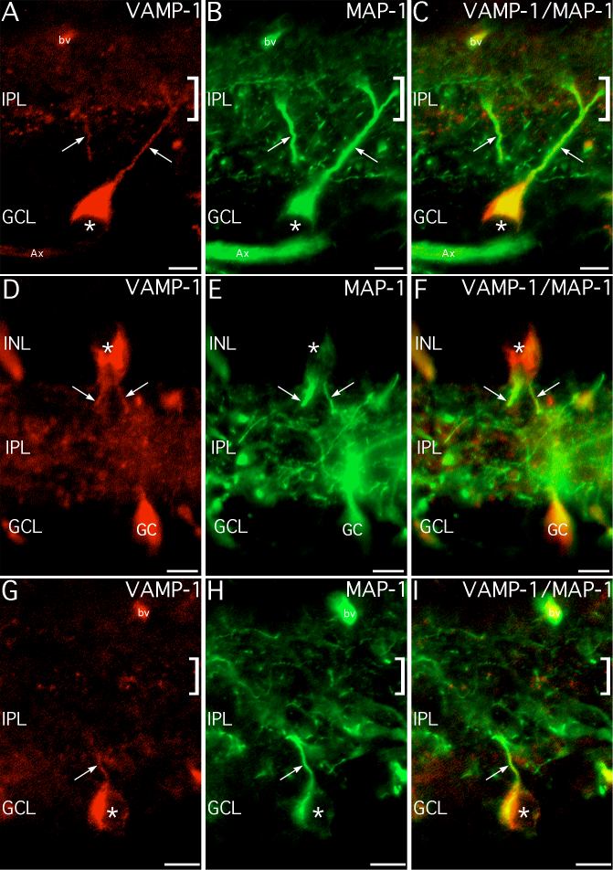
Figure 8. Many VAMP-1-immunoreactive cells are ganglion cells
Double labeling for VAMP-1 and the ganglion cell marker, MAP-1, confirms that many VAMP-1-immunoreactive cells are ganglion cells. A-C: Large VAMP-1-immunoreactive cells (*) with dendrites (arrows) projecting to the VAMP-1-immunoreactive plexus (square bracket symbol) in the mid-IPL show double-labeling for MAP-1, indicating these cells are ganglion cells. A bundle of axons (Ax) showing labeling for both proteins is also visible. Blood vessels (bv) show non-specific labeling. D-F: Large VAMP-1-immunoreactive cells (*) in the INL show double-labeling for MAP-1 in their cell bodies and dendrites (arrows), indicating that they are displaced ganglion cells. A VAMP-1-immunoreactive ganglion cell (GC) double-labeled for MAP-1 is visible in the GCL, but is slightly out of the plane of focus. G-I: Other VAMP-1-immunoreactive cells (*) also show double labeling for MAP-1 in their cell bodies and dendrites (arrow), indicating they are ganglion cells. Blood vessels (bv) show non-specific labeling. Scale bars represent 10 μm.