![]() Figure 7 of
Sherry, Mol Vis 2003;
9:673-688.
Figure 7 of
Sherry, Mol Vis 2003;
9:673-688.
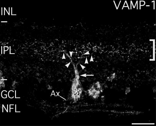
Figure 7. Large VAMP-1 immunoreactive ganglion cells contribute to the VAMP-1 immunoreactive plexus in the mid-IPL
Confocal microscopy shows that the large VAMP-1 immunoreactive ganglion cells contribute to the VAMP-1 immunoreactive plexus in the mid-inner plexiform layer (IPL). Two dimensional projection of a large VAMP-1-immunoreactive ganglion cell (*) in the ganglion cell layer (GCL) constructed from 43 optical sections totaling 12.6 μm thickness. At the level of the mid-IPL the primary dendrite (arrow) splits and smaller branches showing VAMP-1-immunoreactive puncta (arrowheads) distribute in the VAMP-1 plexus in the mid-IPL (square bracket symbol). A fragment of the cell's axon (ax) is also visible, indicating the cell is a ganglion cell. Nearby, part of the cell body of another nearby VAMP-1 immunoreactive cell is visible in the GCL. Several labeled axons are visible in the nerve fiber layer (NFL). INL, inner nuclear layer. Scale bar represents 20 μm.