![]() Figure 3 of
Sherry, Mol Vis 2003;
9:673-688.
Figure 3 of
Sherry, Mol Vis 2003;
9:673-688.
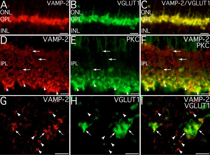
Figure 3. VAMP-2 is expressed at photoreceptor and bipolar cell ribbon synapses
A-C: Double-labeling for VAMP-2 and VGLUT1, a marker for rod and cone terminals in the OPL. Labeling for VAMP-2 colocalizes with labeling for VGLUT1, indicating that VAMP-2 is present in the terminals of rod and cone photoreceptors. D-F: Double-labeling for VAMP-2 and PKC, a marker for rod bipolar cell terminals in the IPL. Labeling for VAMP-2 and PKC colocalizes in the terminals of the rod bipolar cells (arrowheads). Rod bipolar cell axons (arrows) show little labeling for VAMP-2. G-I: Confocal microscopy of double-labeling for VAMP-2 and VGLUT1, a marker for all bipolar cell terminals. Terminals of cone bipolar cells from the mid-IPL are illustrated. Labeling for VAMP-2 colocalizes with VGLUT1, confirming that VAMP-2 is present in cone bipolar cell terminals (arrows) in addition to rod bipolar cells. Several conventional synaptic terminals showing only VAMP-2 labeling (arrowheads) also are present. A single optical section of 0.29 μm thickness is shown. Scale bars represent 10 μm for A-F; 2 μm for G-I.