![]() Figure 12 of
Sherry, Mol Vis 2003;
9:673-688.
Figure 12 of
Sherry, Mol Vis 2003;
9:673-688.
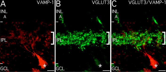
Figure 12. Large VAMP-1-immunoreactive ganglion cells co-stratify with VGLUT3-immunoreactive amacrine cells
The large VAMP-1-immunoreactive ganglion cells co-stratify with VGLUT3-immunoreactive amacrine cells. A: A large VAMP-1-immunoreactive ganglion cell (*) projects to the plexus in the mid-IPL (square bracket symbol). B: The processes of VGLUT3 amacrine cells (A) form a dense plexus that co-stratifies with the VAMP-1 plexus in the mid-IPL (square bracket symbol). C: Overlay of A and B. The VAMP-1 and VGLUT3 plexes in the mid-IPL co-stratify precisely (square bracket symbol). Terminals from the VGLUT3 cells are often seen in close proximity to the VAMP-1-immunoreactive puncta and the dendrites of the large VAMP-1-immunoreactive ganglion cell, suggesting potential synaptic interactions. Scale bar represents 10 μm.