![]() Figure 4 of
Cox, Mol Vis 2003;
9:665-672.
Figure 4 of
Cox, Mol Vis 2003;
9:665-672.
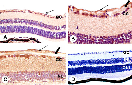
Figure 4. Immunohistochemistry of PDGF-β
Immunohistochemistry of PDGF-β in retinas of diabetic and non-diabetic mice. The Muller cell end-feet at the internal limiting membrane demonstrate most intense immunoreactivity (small arrow; A-C). Lesser DAB reaction product is apparent in the retinal ganglion cells (GC) and inner nuclear layers (INL; A, B) but there is no immunoreactivity in the nerve fiber layer (large arrow) in contrast to PDGF-α localization. There are no clear differences in intensity and staining pattern between non-diabetic (A, B) and diabetic retinas (C). Controls showed no immunoreactivity (D). Original magnifications: x200 (A, D); x400 (B, C). The outer nuclear layer (ONL) is also labeled in the micrograph.