![]() Figure 3 of
Cox, Mol Vis 2003;
9:665-672.
Figure 3 of
Cox, Mol Vis 2003;
9:665-672.
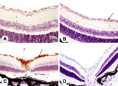
Figure 3. Immunohistochemistry of PDGF-α
Immunohistochemistry of PDGF-α in retinas of diabetic and non-diabetic mice. The retinal ganglion cells (GC) and nerve fiber layer (arrow) of both non-diabetic (A) and diabetic (B) show strong immunoreactivity. The axonal concentration of this receptor (far removed from the cell body) is exemplified by strong PDGF-α immunreactivity at the optic nerve head where ganglion cell axons coalesce (C, arrow). Controls show no apparent deposition of DAB reaction product (D). Original magnifications: x200 (A, B); x100 (C, D). The outer nuclear layer (ONL) and the inner nuclear layer (INL) are also labeled in the micrograph.