![]() Figure 5 of
Du, Mol Vis 2003;
9:635-643.
Figure 5 of
Du, Mol Vis 2003;
9:635-643.
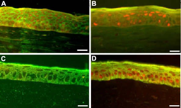
Figure 5. Grafted human cells in transplanted rabbit corneas
A and B: Immunofluorescent staining of corneas with HAM-LC transplantation. C and D: Normal rabbit cornea staining. A and C show double staining with anti-human nuclear antibody (positive shows red in nucleus) and K3 (positive shows green in cytoplasm). B and D show double staining with p63 (red in nucleus) and K3. The bars represent 20 μm.