![]() Figure 4 of
Du, Mol Vis 2003;
9:635-643.
Figure 4 of
Du, Mol Vis 2003;
9:635-643.
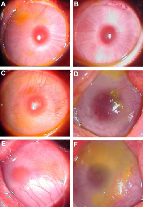
Figure 4. Reconstruction of rabbit corneas with LSCD by cultured limbal cell transplantation
The illustrations show the ocular surface of rabbits pre- (left panels) and post-operation (right panels). A, C, and E show rabbits with LSCD before transplantation, illustrating corneal vascularization and epithelial defects by fluorescein staining. B shows forty days after transplantation of human limbal stem cells cultured on HAM to the eye shown in A. D shows forty days after amniotic membrane transplantation to the eye in C. F shows forty days after keratectomy alone to the eye in E, without transplantation. Transplanted eyes were covered with HAM to 2 mm beyond the visible limbal area.