![]() Figure 3 of
Du, Mol Vis 2003;
9:635-643.
Figure 3 of
Du, Mol Vis 2003;
9:635-643.
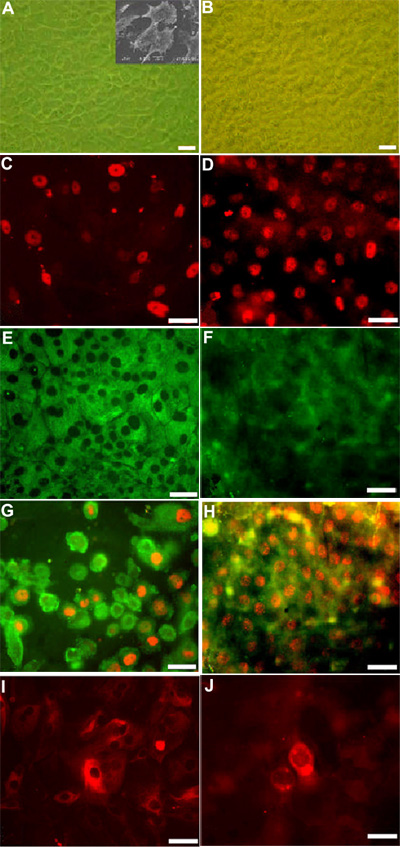
Figure 3. Cells cultured on HAM retain markers of progenitor cells better than those cultured on plastic
Limbal cells were cultured on plastic (left panels) or on HAM (right panels). Cells cultured on plastic (A) form a monolayer with more uniform morphology and denser arrangement than on HAM (B). Inset in A shows electron microscopic characteristics of cultured limbal cells. C and D are TRITC-conjugated p63 nuclear staining. E and F are connexin43 staining. G and H are double-stained with p63 and connexin43. I and J are TRITC-conjugated antibody to K3. The bars represent 10 μm.