![]() Figure 1 of
Du, Mol Vis 2003;
9:635-643.
Figure 1 of
Du, Mol Vis 2003;
9:635-643.
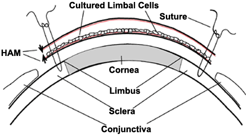
Figure 1. Transplantation of cultured limbal cells onto LSCD rabbit corneas
As described under Methods, one month after ablation of corneal epithelial and limbal cells, the covering conjunctival cellular material was removed by scraping from a region 5 mm beyond the Limbus. Human amniotic membrane (HAM) carrying cultured human limbal cells on the basement membrane surface (colored) was sutured on the denuded region and covered by a second HAM with the basement membrane surface oriented toward the cornea. Orientation of the HAM basement membrane is shown by Arrows.