![]() Figure 2 of
Lee, Mol Vis 2003;
9:624-634.
Figure 2 of
Lee, Mol Vis 2003;
9:624-634.
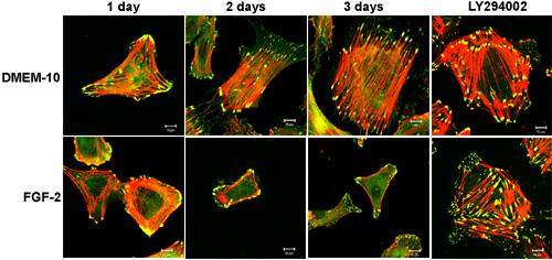
Figure 2. Immunofluorescent staining of CECs with phalloidin and anti-vinculin antibody
First passage (1x10⁴) cells were plated in a 4 well chamber and maintained for 2 days in the presence or absence of 10 ng/ml of FGF-2 in DMEM-10. On day 3, cells were further maintained in DMEM-10 or FGF-2 or were simultaneously treated with FGF-2 and 20 μM LY294002 for an additional 24 h. Cells were fixed and stained for vinculin (green) and F-actin (red) and merged images are shown in yellow. Data shown are representative of four experiments. Bar represents 10 μm.