![]() Figure 1 of
Lee, Mol Vis 2003;
9:624-634.
Figure 1 of
Lee, Mol Vis 2003;
9:624-634.
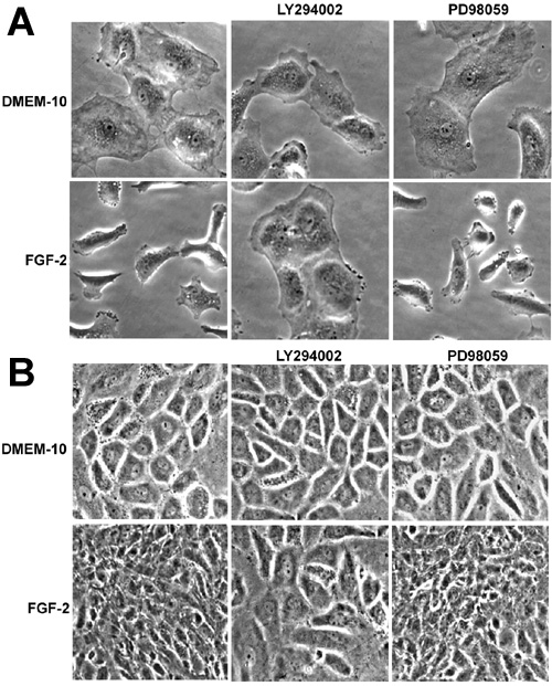
Figure 1. FGF-2 mediated cell shape change of CECs adherent to tissue culture substratum
First passage cells were plated in 35 mm dishes at a cell density of 1x10⁵ (A) or 3x10⁵ (B) in DMEM-10 for 2 days (control cells). Other first passage cells were treated with 10 ng/ml of FGF-2 in DMEM-10 for 2 days. Thereafter, cells were treated with either 20 μM of LY294002 or 10 μM of PD98059 for an additional 24 h. Phase contrast micrographs were then taken with a digital camera. Data shown are representative of four experiments. All images were magnified 200 times from their original size.